Ventricular Septal Defect Vsd American Heart Association

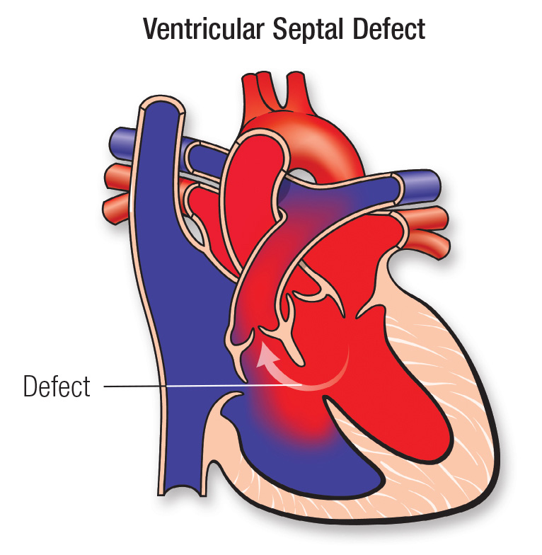
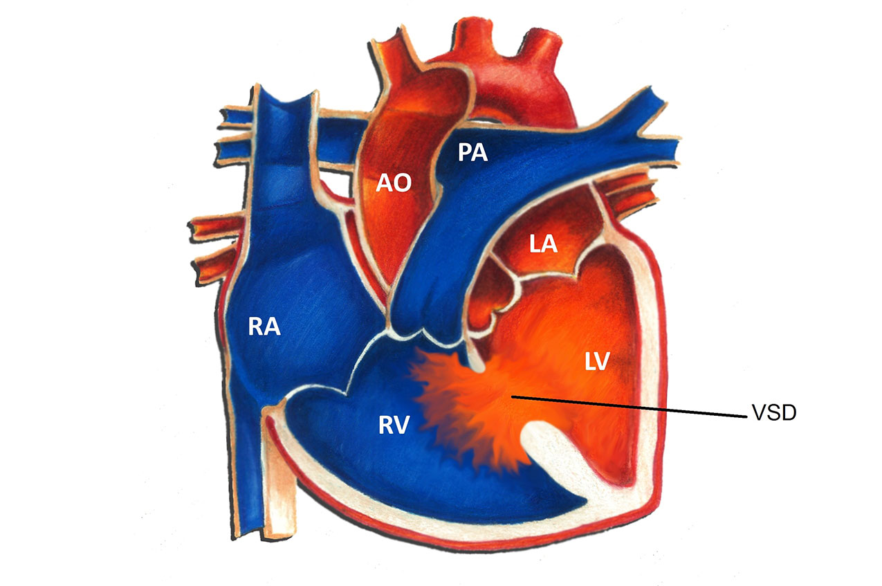
Ventricular Septal Defect Vsd American Heart Association Vsd is an opening or hole (defect) in the wall (septum) separating the two lower chambers of the heart (ventricles). in normal development, the wall between the chambers closes before the fetus is born, so that by birth, oxygen rich blood is kept from mixing with the oxygen poor blood. when the hole does not close, it may cause higher pressure. The present article describes the clinical aspects of ventricular septal defects and current management strategies. ventricular septal defect (vsd) is a common congenital heart defect in both children and adults. management of this lesion has changed dramatically in the last 50 years. catheter based therapy for vsd closure, now in the clinical.

Ventricular Septal Defect Vsd Pediatric Heart Specialists Key search words included but were not limited to the following: adult congenital heart disease, anesthesia, aortic aneurysm, aortic stenosis, atrial septal defect, arterial switch operation, bradycardia, bicuspid aortic valve, cardiac catheterization, cardiac imaging, cardiovascular magnetic resonance, cardiac reoperation, cardiovascular. The american heart association explains the common types of congenital defects including aortic valve stenosis, avs, atrial septal defect, asd, coarctation of the aorta, coa, complete atrioventricular canal defect, cavc, d transposition of the great arteries, ebstein's anomaly, i transposition of the great arteries, patent ductus arteriosis, pda, pulmonary valve stenosis, single ventricle. Ventricular septal defects (vsds) are the most common congenital cardiac anomaly in children and are the second most common congenital abnormality in adults, surpassed only by a bicuspid aortic valve. the primary pathophysiology involves an abnormal communication between the right and left ventricles, leading to shunt formation and subsequent hemodynamic compromise in vsd. while spontaneous. Survival depends on there being an opening in the wall between the atria (atrial septal defect) and usually an opening in the wall between the two ventricles (ventricular septal defect). as a result, the low oxygen blood that returns from the body veins to the right atrium flows through the atrial septal defect and into the left atrium.

Comments are closed.