Ventricular Septal Defect Etiology Pathophysiology Clinical

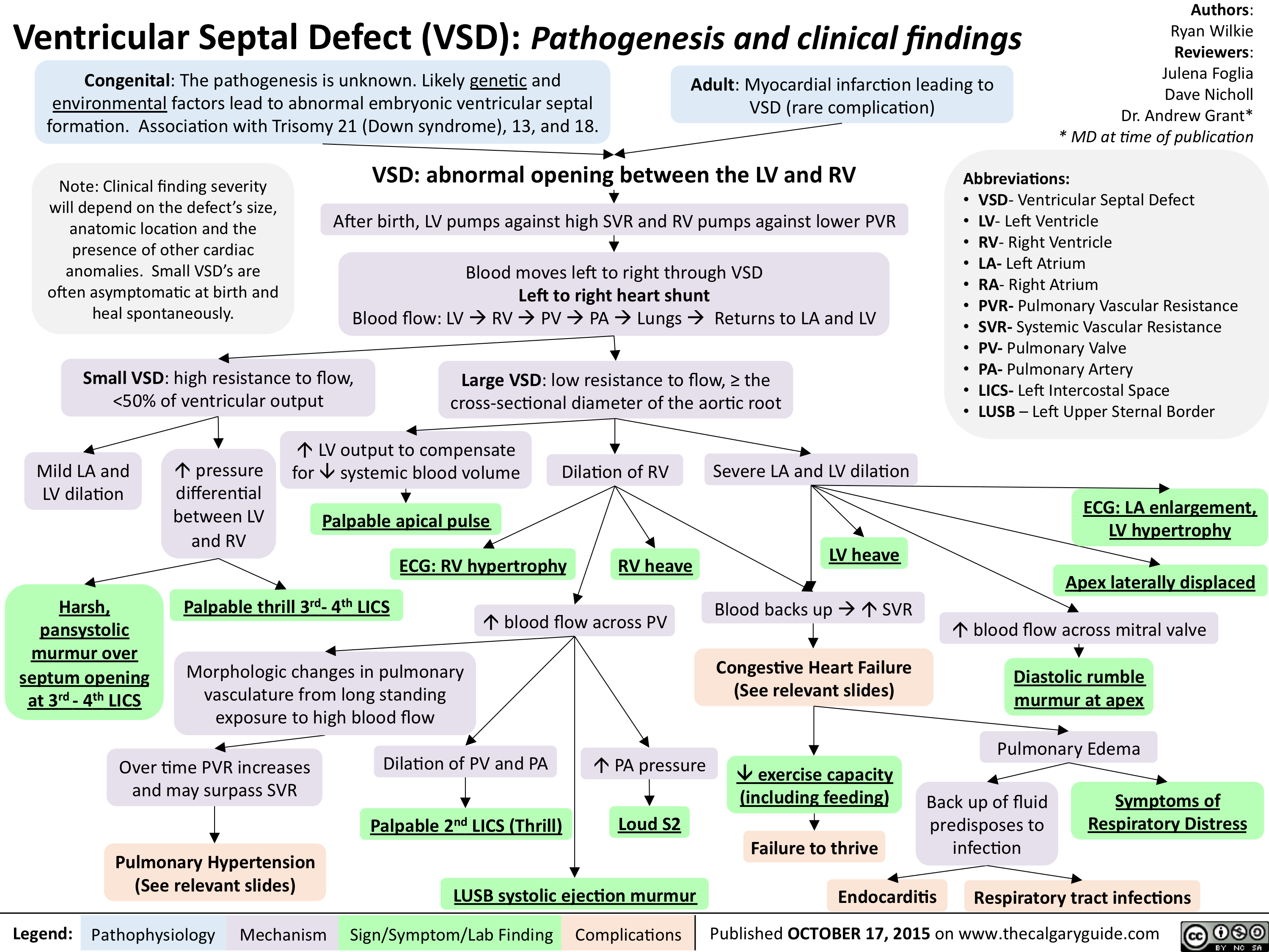
Ventricular Septal Defect Vsd American Heart Association Ventricular septal defects (vsds) are the most common congenital cardiac anomaly in children and are the second most common congenital abnormality in adults, surpassed only by a bicuspid aortic valve. the primary pathophysiology involves an abnormal communication between the right and left ventricles, leading to shunt formation and subsequent hemodynamic compromise in vsd. while spontaneous. A ventricular septal defect (vsd) is a hole in the heart that's present at birth (congenital heart defect). the hole is between the lower heart chambers (right and left ventricles). it allows oxygen rich blood to move back into the lungs instead of being pumped to the rest of the body. a ventricular septal defect (vsd) changes how blood flows.

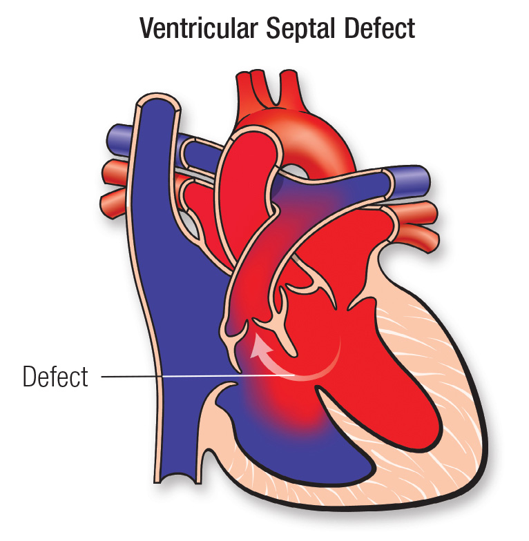
Ventricular Septal Defect Vsd Pathogenesis And Clinical Findings Ventricular septal defect (vsd) is one of the most common congenital heart defects (second only to bicuspid aortic valve) at birth, but accounts for only 10 percent of congenital heart defects in adults because many close spontaneously [1,2]. vsds are of various sizes and locations, can be single or multiple, and may occur as isolated lesions. A ventricular septal defect is a hole in the wall that separates the lower chambers of your heart. when this hole is large enough, the amount of blood leaking between the chambers can cause permanent damage to your heart and lungs and increase the risk of heart infections. most vsds don’t cause symptoms and close on their own by age 6. Ventricular septal defect (see figure ventricular septal defect) is the 2nd most common congenital heart anomaly after bicuspid aortic valve, accounting for 20% of all defects. it can occur alone or with other congenital anomalies (eg, tetralogy of fallot , complete atrioventricular septal defects , transposition of the great arteries ). The present article describes the clinical aspects of ventricular septal defects and current management strategies. ventricular septal defect (vsd) is a common congenital heart defect in both children and adults. management of this lesion has changed dramatically in the last 50 years. catheter based therapy for vsd closure, now in the clinical.

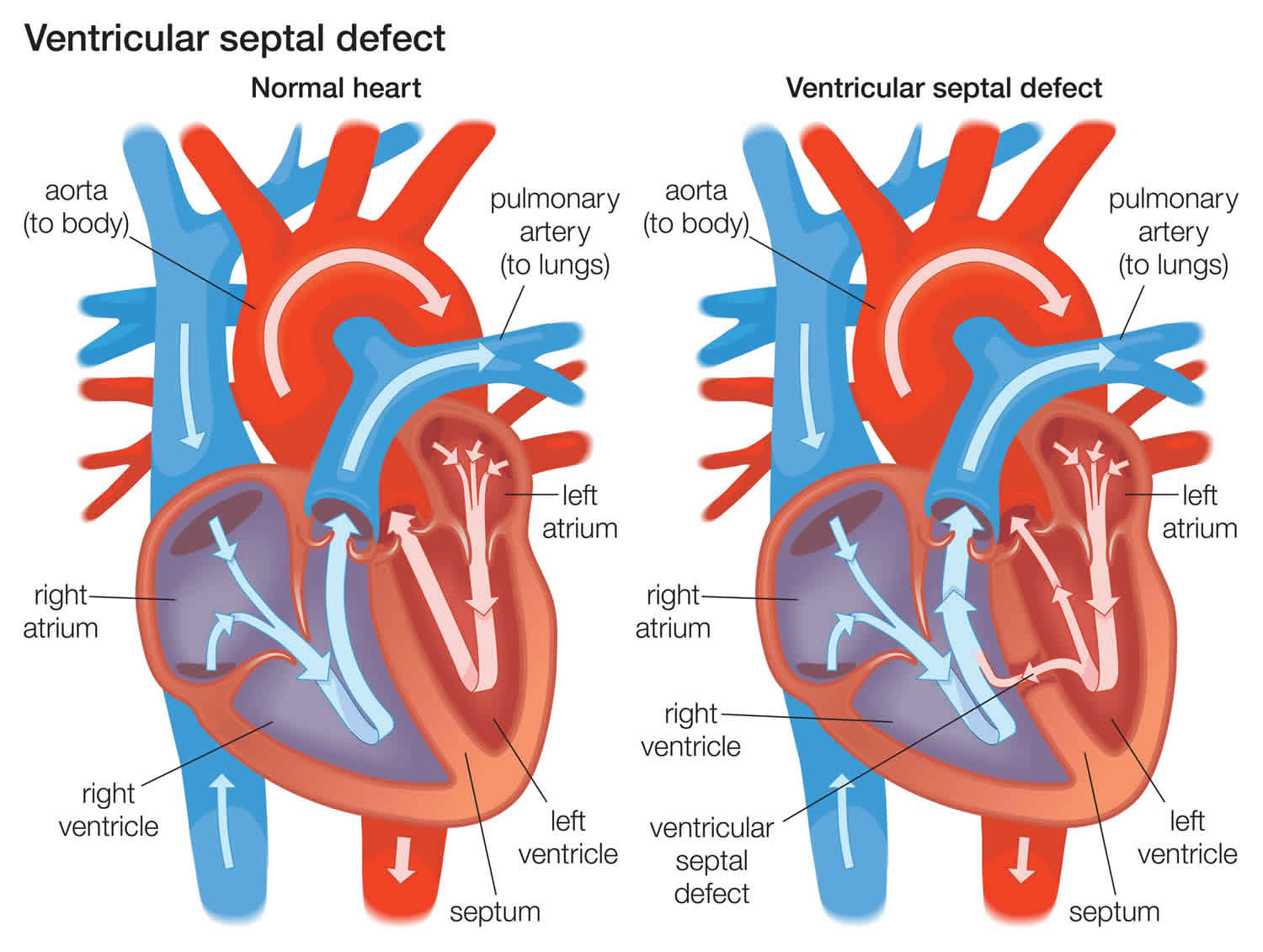
Ventricular Septal Defect Causes Types Symptoms Diagnosis Treatment Ventricular septal defect (see figure ventricular septal defect) is the 2nd most common congenital heart anomaly after bicuspid aortic valve, accounting for 20% of all defects. it can occur alone or with other congenital anomalies (eg, tetralogy of fallot , complete atrioventricular septal defects , transposition of the great arteries ). The present article describes the clinical aspects of ventricular septal defects and current management strategies. ventricular septal defect (vsd) is a common congenital heart defect in both children and adults. management of this lesion has changed dramatically in the last 50 years. catheter based therapy for vsd closure, now in the clinical. A ventricular septal defect (vsd) is a hole or a defect in the septum that divides the two lower chambers of the heart, resulting in communication between the ventricular cavities. a vsd may occur as a primary anomaly, with or without additional major associated cardiac defects. it may also occur as a single component of a wide variety of. The heart is made up of four chambers. a ventricular septal defect is an abnormal opening on the wall (or septum) that separates the heart’s two lower chambers, or ventricles, which are also the blood’s main pumping chambers. in a person with a normal heart, blood moves from the heart’s right ventricle to the lungs, where it is enriched.

Ventricular Septal Defect Etiology Pathophysiology Clinical A ventricular septal defect (vsd) is a hole or a defect in the septum that divides the two lower chambers of the heart, resulting in communication between the ventricular cavities. a vsd may occur as a primary anomaly, with or without additional major associated cardiac defects. it may also occur as a single component of a wide variety of. The heart is made up of four chambers. a ventricular septal defect is an abnormal opening on the wall (or septum) that separates the heart’s two lower chambers, or ventricles, which are also the blood’s main pumping chambers. in a person with a normal heart, blood moves from the heart’s right ventricle to the lungs, where it is enriched.

Comments are closed.