Ventricular Septal Defect Clinical Gate

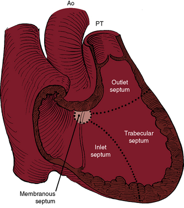
Ventricular Septal Defect Clinical Gate A vsd is a defect in the interventricular septum. vsds may occur anywhere, but are most commonly found at points of junction of the embryologic components of the septum (i.e., the main [or posterior] ventricular septum, the bulbar [or infundibular] septum, and the membranous septum). by dividing the septum into four components (membranous. The tendency for ventricular septal defects, especially perimembranous and trabecular muscular defects, to decrease in size finds an ultimate expression in complete closure 24,31–33 that has been called the therapeutics of nature—the invisible sutures of spontaneous closure. 34 the incidence rate of spontaneous closure varies considerably.

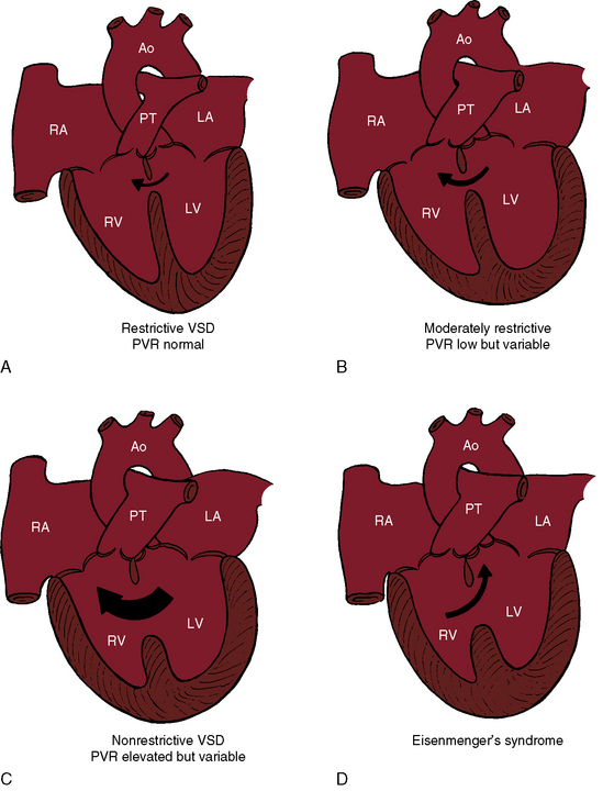
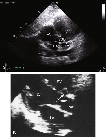
Ventricular Septal Defect Clinical Gate There is a moderate (5 mm) perimembranous ventricular septal defect (vsd) (subaortic), with bidirectional shunting and velocity of 1 m s . 4. outflow assessment shows normally related great arteries with normal valve morphology and size. the pulmonary artery is anterior to and to the left of the aorta. 5. Ventricular septal defects (vsds) are the most common congenital cardiac anomaly in children and are the second most common congenital abnormality in adults, surpassed only by a bicuspid aortic valve. the primary pathophysiology involves an abnormal communication between the right and left ventricles, leading to shunt formation and subsequent hemodynamic compromise in vsd. while spontaneous. A ventricular septal defect (vsd) is a hole or defect. in the s eptum that separates the heart's two bottom. chambers and allows communication between them. a vsd can occur as a single anomaly or. Ventricular septal defect (vsd) is one of the most common congenital heart defects (second only to bicuspid aortic valve) at birth, but accounts for only 10 percent of congenital heart defects in adults because many close spontaneously [1,2]. vsds are of various sizes and locations, can be single or multiple, and may occur as isolated lesions.

Ventricular Septal Defect Clinical Gate A ventricular septal defect (vsd) is a hole or defect. in the s eptum that separates the heart's two bottom. chambers and allows communication between them. a vsd can occur as a single anomaly or. Ventricular septal defect (vsd) is one of the most common congenital heart defects (second only to bicuspid aortic valve) at birth, but accounts for only 10 percent of congenital heart defects in adults because many close spontaneously [1,2]. vsds are of various sizes and locations, can be single or multiple, and may occur as isolated lesions. Ventricular septal defect (see figure ventricular septal defect) is the 2nd most common congenital heart anomaly after bicuspid aortic valve, accounting for 20% of all defects. it can occur alone or with other congenital anomalies (eg, tetralogy of fallot , complete atrioventricular septal defects , transposition of the great arteries ). A ventricular septal defect (vsd) is a hole in the heart that's present at birth (congenital heart defect). the hole is between the lower heart chambers (right and left ventricles). it allows oxygen rich blood to move back into the lungs instead of being pumped to the rest of the body. a ventricular septal defect (vsd) changes how blood flows.

Comments are closed.