Vena Cavae Anatomy Of The Superior Vena Cava And Inferior Vena Cava

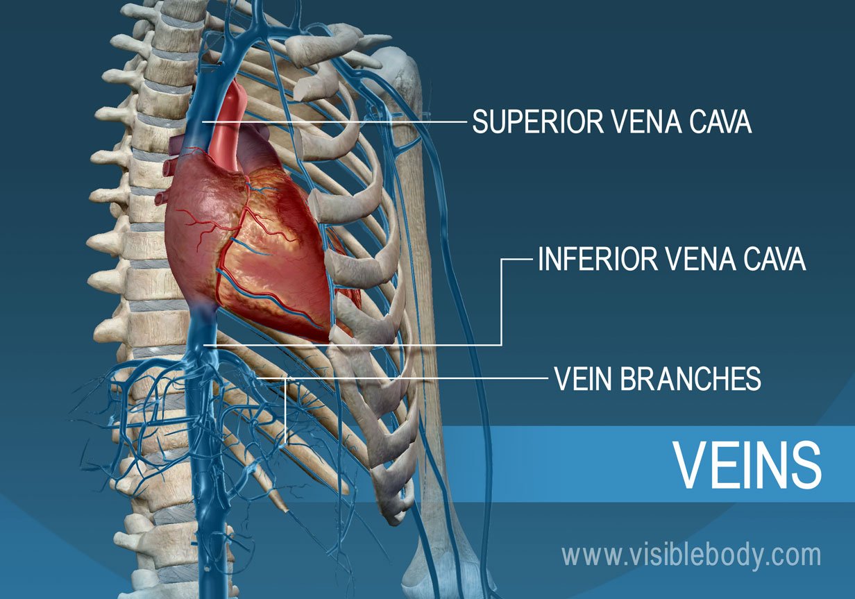
Difference Between Superior And Inferior Vena Cava Pediaa Com Your superior vena cava and inferior vena cava have the important function of carrying oxygen poor blood to your heart’s right atrium, where it moves into your right ventricle and then to your lungs (through your pulmonary artery) to trade in carbon dioxide for oxygen. oxygenated blood comes back through your pulmonary veins to your heart’s. The superior vena cava (svc) is one of the two largest veins in the body. it is a vital part of the cardiovascular system, responsible for returning deoxygenated blood, or blood that is rich in carbon dioxide, from the upper body to the heart. the superior vena cava has thin walls that render it vulnerable to high blood pressure.

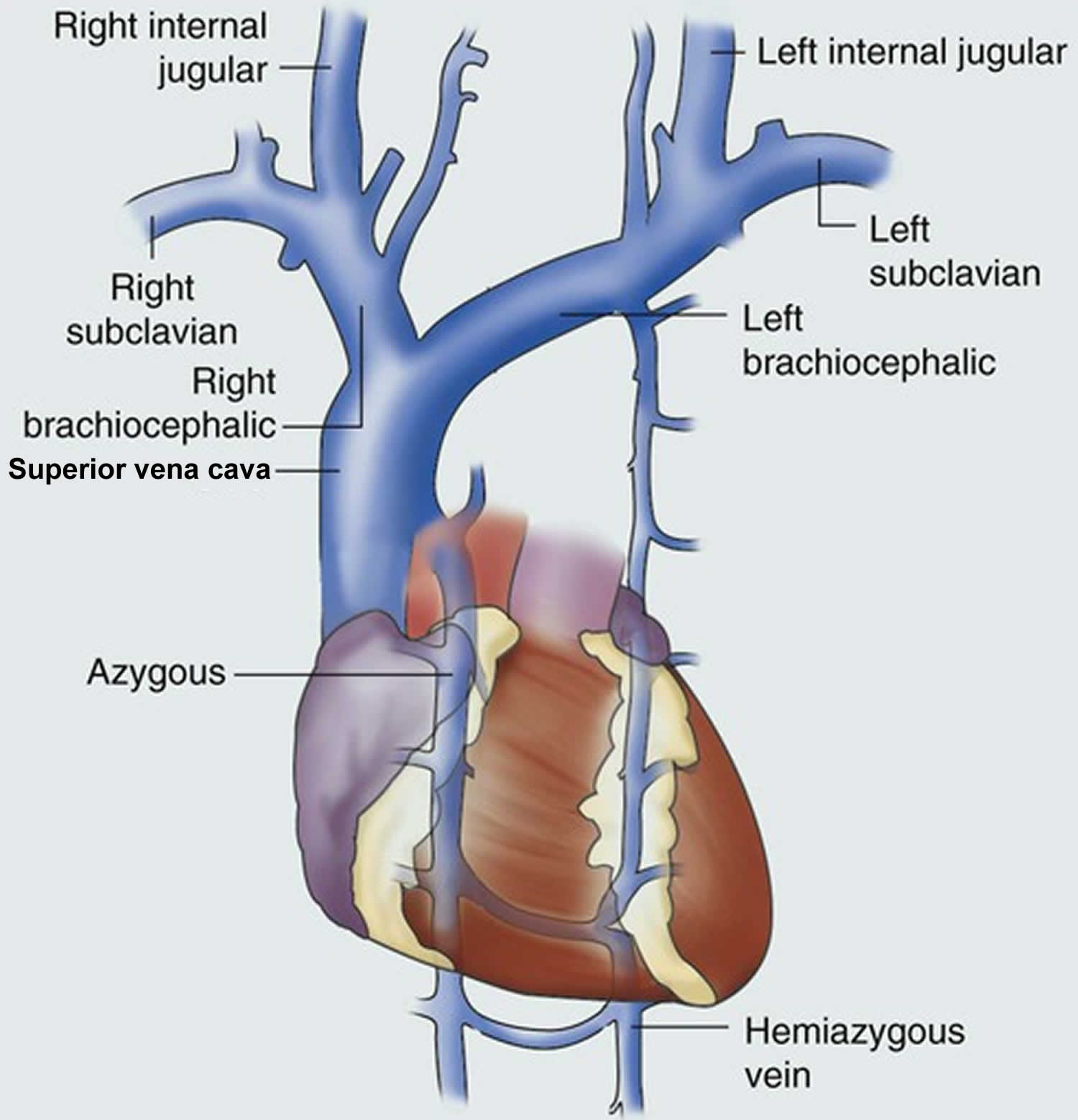
Superior Vena Cava Anatomy Function Superior Vena Cava Syn Anatomy and function of the superior vena cava. the superior vena cava (svc, also known as the cava or cva) is a short, but large diameter vein located in the anterior right superior mediastinum. its latin name is related to its large pipe appearance in cadavers, 'cava' meaning 'hollow'. the superior vena cava is very important for the function. The superior vena cava is located in the upper chest region and is formed by the joining of the brachiocephalic veins. these veins drain blood from the upper body regions including the head, neck, and chest. it is bordered by heart structures such as the aorta and pulmonary artery. the inferior vena cava is formed by the joining of the common. The superior vena cava (svc) is a large, significant vein responsible for returning deoxygenated blood collected from the body to the right atrium. it is present within the superior and middle mediastinum. the superior vena cava handles the venous return of blood from structures located superior to the diaphragm. in contrast, its counterpart, the inferior vena cava, handles venous return from. The superior vena cava is a thin walled, low pressure vessel which makes it vulnerable to compression. superior vena cava obstruction can occur either due to external compression or from an occlusion within the vessel lumen itself. the most common cause of svc obstruction is malignancy, typically from lung cancer, lymphoma, or metastatic disease.

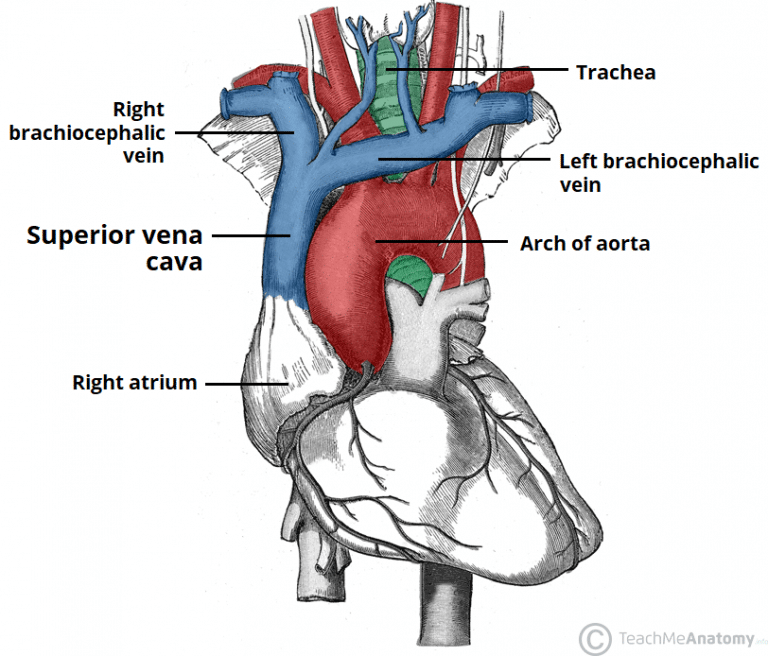
The Superior Vena Cava Teachmeanatomy The superior vena cava (svc) is a large, significant vein responsible for returning deoxygenated blood collected from the body to the right atrium. it is present within the superior and middle mediastinum. the superior vena cava handles the venous return of blood from structures located superior to the diaphragm. in contrast, its counterpart, the inferior vena cava, handles venous return from. The superior vena cava is a thin walled, low pressure vessel which makes it vulnerable to compression. superior vena cava obstruction can occur either due to external compression or from an occlusion within the vessel lumen itself. the most common cause of svc obstruction is malignancy, typically from lung cancer, lymphoma, or metastatic disease. The superior vena cava transports blood from the head, neck, upper limbs and thorax into the right atrium. the svc is approximately 7cm long and does not have any valves. it is located in the right superior mediastinum. the svc is formed by the right and left brachiocephalic veins joining together just behind the lower border of the first. The superior vena cava is a large (measures .78 inches in diameter and 2.7 inches in length. [4]), significant vein responsible for returning deoxygenated blood collected from the body back into the heart. the superior vena cava starts at the lower border of the first costal cartilage. it is located posterior (behind) this first costal.

Blood Vessels Circulatory Anatomy The superior vena cava transports blood from the head, neck, upper limbs and thorax into the right atrium. the svc is approximately 7cm long and does not have any valves. it is located in the right superior mediastinum. the svc is formed by the right and left brachiocephalic veins joining together just behind the lower border of the first. The superior vena cava is a large (measures .78 inches in diameter and 2.7 inches in length. [4]), significant vein responsible for returning deoxygenated blood collected from the body back into the heart. the superior vena cava starts at the lower border of the first costal cartilage. it is located posterior (behind) this first costal.

Comments are closed.