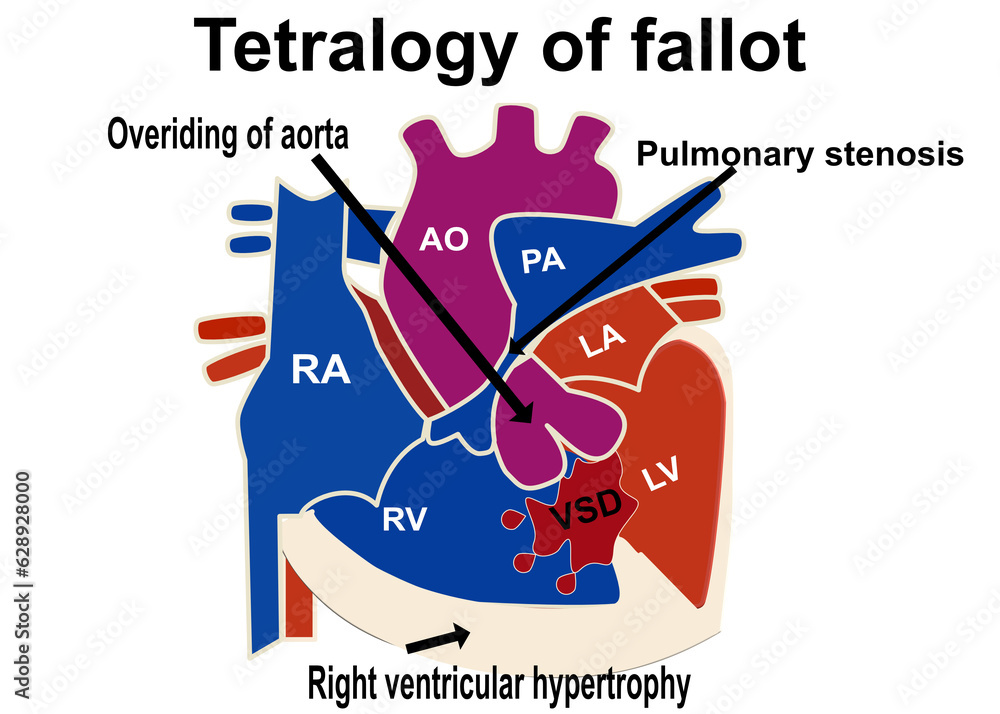
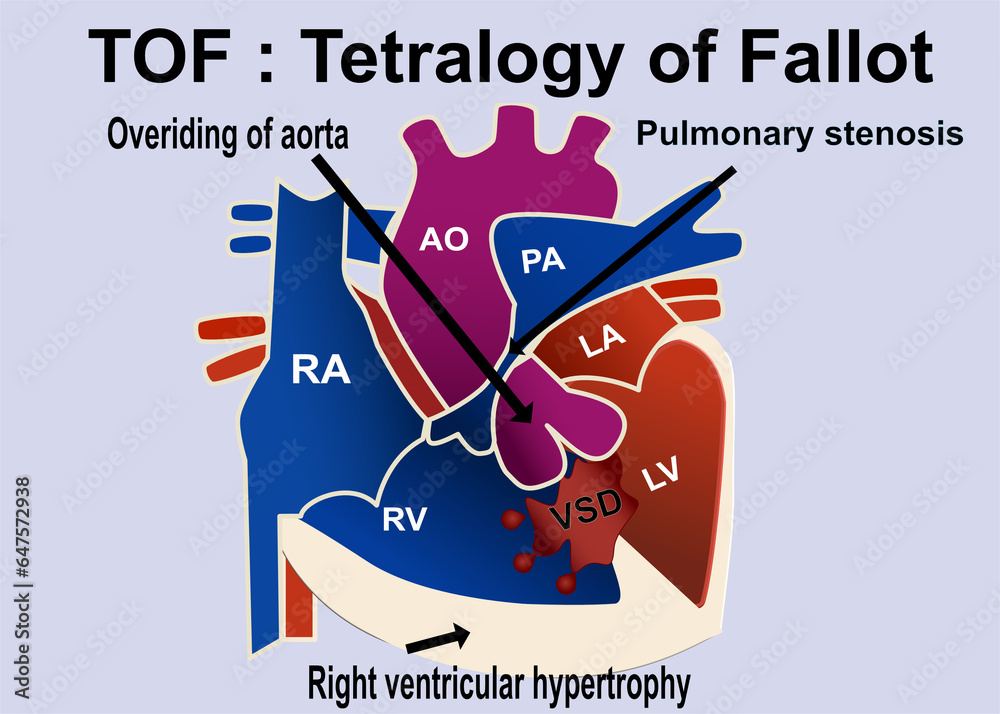
The Picture Show The Structure Of Tetralogy Of Fallot That Show The 4

Tetralogy Of Fallot American Heart Association This test can show problems with the structure of the heart and how the heart is working with this defect. after the baby is born. tetralogy of fallot usually is diagnosed after a baby is born, often after the infant has an episode of turning blue during crying or feeding (a tet spell). Tetralogy of fallot occurs as the baby's heart grows during pregnancy. usually, the cause is unknown. tetralogy of fallot includes four problems with heart structure: narrowing of the valve between the heart and the lungs, called pulmonary valve stenosis. this condition reduces blood flow from the heart to the lungs.

The Picture Show The Structure Of Tetralogy Of Fallot That Show The 4 Tetralogy of fallot is treated with two kinds of surgery. one provides temporary improvement by a shunt to give more blood flow to the lungs. the other is a complete repair of the two most important abnormalities that make up tetralogy of fallot. patients might have one or both surgeries in their lifetime. 2. ventricular septal defect. with significant obstruction in the right ventricular outflow tract, blood will shunt from right to left, bypassing the lungs and leading to cyanosis. 3. right ventricular hypertrophy (thickened muscle wall) secondary to higher pressure load on this chamber. 4. Tetralogy of fallot is a heart condition you’re born with that makes it hard to get enough oxygen to your body because of four abnormalities in your heart’s structure. surgery in infancy repairs the issues and helps blood flow better, but you’ll need lifelong follow ups with a provider. Classic tetralogy of fallot (tof) is a congenital heart defect (chd) that is comprised of 4 anatomical alterations: a large, anteriorly malaligned ventricular septal defect (vsd), an overriding aorta which results in infundibular (ie, sub pulmonary) right ventricular outflow tract obstruction (rvoto), and consequent right ventricular hypertrophy secondary to chronic systemic pressures. the.

Tetralogy Of Fallot Pathophysiology Wikidoc Tetralogy of fallot is a heart condition you’re born with that makes it hard to get enough oxygen to your body because of four abnormalities in your heart’s structure. surgery in infancy repairs the issues and helps blood flow better, but you’ll need lifelong follow ups with a provider. Classic tetralogy of fallot (tof) is a congenital heart defect (chd) that is comprised of 4 anatomical alterations: a large, anteriorly malaligned ventricular septal defect (vsd), an overriding aorta which results in infundibular (ie, sub pulmonary) right ventricular outflow tract obstruction (rvoto), and consequent right ventricular hypertrophy secondary to chronic systemic pressures. the. Learn more about the cardiac center. outpatient appointments. 215 590 4040. second opinions, referrals and information about our services. 267 426 9600. general inquiries. refer a patient. children’s hospital of philadelphia's pediatric heart surgery survival rates are among the best in the nation. learn more. Anatomy and pathophysiology of tetralogy of fallot (tof): normal heart structure (a) promotes unidirectional flow of deoxygenated blood (blue) into the lungs and oxygenated blood (red) into the aorta; in tof (b) pulmonary stenosis and narrowing of the right ventricular outflow tract (rvot) impedes the flow of deoxygenated blood into the lungs, and both the ventricular septal defect (vsd) and.

The Picture Show The Structure Of Tetralogy Of Fallot That Show The 4 Learn more about the cardiac center. outpatient appointments. 215 590 4040. second opinions, referrals and information about our services. 267 426 9600. general inquiries. refer a patient. children’s hospital of philadelphia's pediatric heart surgery survival rates are among the best in the nation. learn more. Anatomy and pathophysiology of tetralogy of fallot (tof): normal heart structure (a) promotes unidirectional flow of deoxygenated blood (blue) into the lungs and oxygenated blood (red) into the aorta; in tof (b) pulmonary stenosis and narrowing of the right ventricular outflow tract (rvot) impedes the flow of deoxygenated blood into the lungs, and both the ventricular septal defect (vsd) and.

Comments are closed.