Ppt Dental X Ray Safety Training Powerpoint Presentation Free

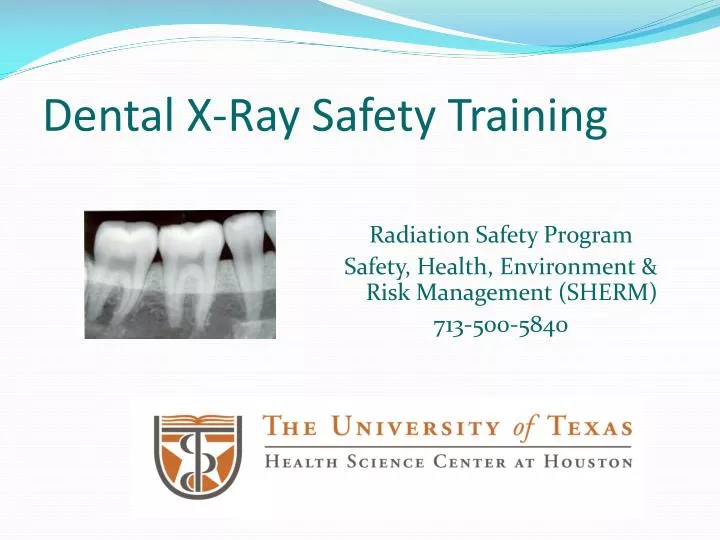
Ppt Dental X Ray Safety Training Powerpoint Presentation Free 1) licensed dentists must maintain radiation exposures as low as reasonably achievable and understand the health risks of radiation. 2) dental radiographic equipment must be registered and follow safety protocols to protect patients and staff, such as using protective gear and collimation. 3) dentists are responsible for quality assurance. Alara techniques • minimize the time spent in unshielded environments while the x ray beam is on • avoid anyone beyond the patient being in the area while x ray beam is on • stand at least 6 feet away from the dental x ray machine • standing 6ft away verses 1 ft away reduces the exposure by 1 36. alara techniques • x rays are easily.

Ppt Dental X Ray Safety Powerpoint Presentation Free Downl Panoramic the panoramic x ray is used to check on wisdom teeth, assess jaw problems and plan for dental implants. this x ray rotates around the head. periapical the periapical x ray looks at 1 2 teeth at a time. it highlights the entire length of the tooth from the root to the crown. Dental radiography involves taking images of the teeth, bones, and soft tissues in the mouth to aid in diagnosis and treatment planning. there are several types of dental radiography procedures, including intraoral radiographs like bitewings and periapicals, as well as panoramic and cephalometric images. radiographs are useful for detecting. Presentation transcript. dental radiography safety by aggie barlow, chp, ms, mba. goal the goal of dental radiography is to obtain useful diagnostic information while keeping radiation exposure to the patient and dental staff to a minimum. digital radiography in dentistry digital radiography was introduced in dentistry in 1987. Dec 23, 2010 • download as ppt, pdf •. 30 likes • 14,848 views. ai enhanced description. smile care. dental x rays help visualize parts of the teeth and jaws that cannot be seen normally. there are different types of intra oral and extra oral x rays used for various purposes like detecting decay, evaluating bone quality, and examining the.

Ppt Dental X Ray Safety Training Powerpoint Presentation Free Presentation transcript. dental radiography safety by aggie barlow, chp, ms, mba. goal the goal of dental radiography is to obtain useful diagnostic information while keeping radiation exposure to the patient and dental staff to a minimum. digital radiography in dentistry digital radiography was introduced in dentistry in 1987. Dec 23, 2010 • download as ppt, pdf •. 30 likes • 14,848 views. ai enhanced description. smile care. dental x rays help visualize parts of the teeth and jaws that cannot be seen normally. there are different types of intra oral and extra oral x rays used for various purposes like detecting decay, evaluating bone quality, and examining the. Instantly convert your powerpoint training to mobile – for free. experience the magic of sc training (formerly edapp) on your own training content. upload your powerpoint file and our powerful ai doc transformer (coming soon) will instantly make it mobile friendly. only .pptx files accepted. maximum file size 100mb. Download ppt "introduction to dental radiography and equipment". discovery of the x ray roentgen 1895 walkoff kells anode cathode dental radiograph kells intraoral radiograph william conrad roentgen discovered x rays in 1895 by using a glass vacuum tube with an electrical circuit connected to each end. the stream of electrons traveled from the.

Comments are closed.