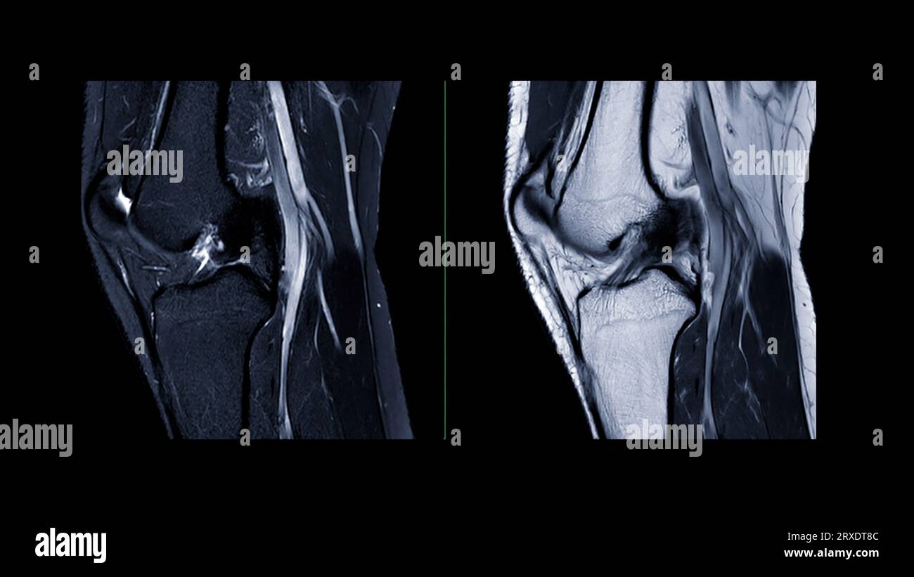
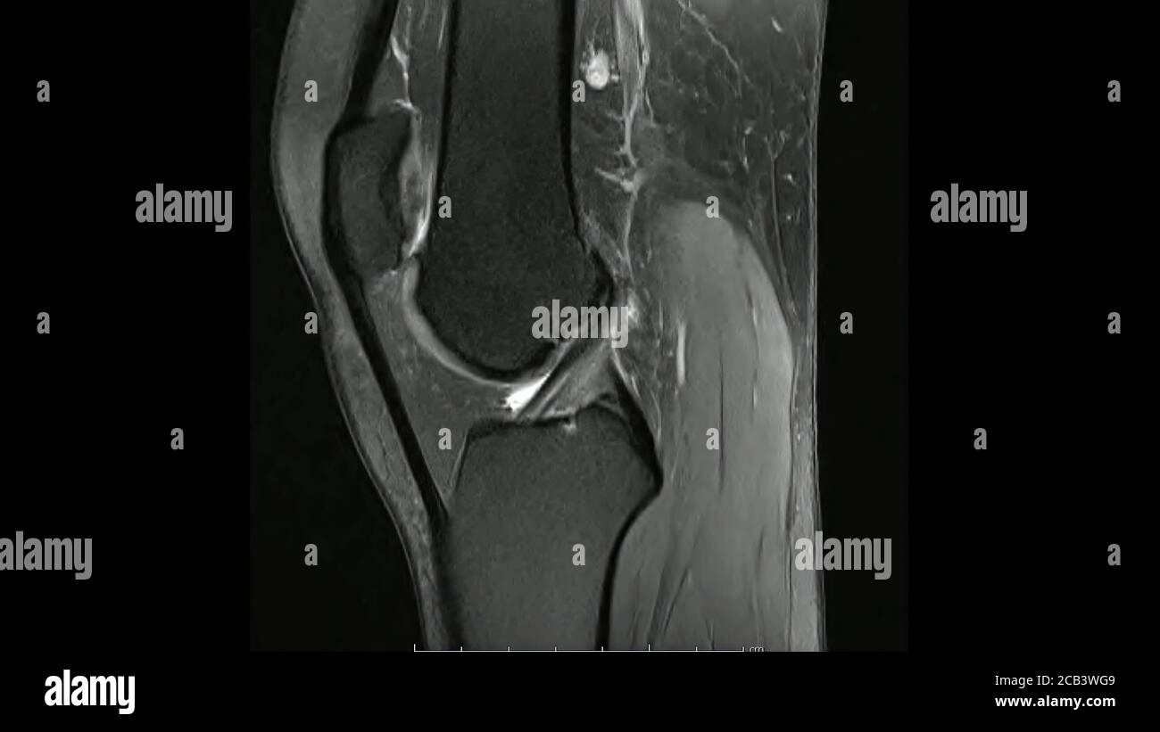
Magnetic Resonance Image Of The Knee Joint Mri Kneein Sagitta

Magnetic Resonance Image Of The Knee Joint Mri Kneein Sagi Abstract. knee pain is frequently seen in patients of all ages, with a wide range of possible aetiologies. magnetic resonance imaging (mri) of the knee is a common diagnostic examination performed for detecting and characterising acute and chronic internal derangement injuries of the knee and helps guide patient management. Magnetic resonance (mr) imaging is the most important imaging modality for the evaluation of traumatic or degenerative cartilaginous lesions in the knee. it is a powerful noninvasive tool for detecting such lesions and monitoring the effects of pharmacologic and surgical therapy. the specific mr imaging techniques used for these purposes can be divided into two broad categories according to.

Knee Joint Coloured Magnetic Resonance Image Mri Of A Sagittal Mri based knee investigation is usually performed on joints at rest or in a non weight bearing condition that does not mimic the actual physiological condition of the joint. this discrepancy may lead to missed detections of early stage oa or of minor lesions. the mechanical properties of degenerated musculoskeletal (msk) tissues may vary. Magnetic resonance imaging (mri) of the knee is a common diagnostic examination performed for detecting and characterising acute and chronic internal derangement injuries of the knee and helps guide patient management. this article reviews the current clinical practice of mri evaluation and interpretation of meniscal, ligamentous, cartilaginous. Magnetic resonance imaging (mri) is one of the most widely used investigations for knee pain as it provides detailed assessment of the bone and soft tissues. the aim of this study is to report the frequency of each diagnosis identified on mri scans of the knee and explore the relationship between mri results and onward treatment. Magnetic resonance imaging (mri) of the knee uses a powerful magnetic field, radio waves and a computer to produce detailed pictures of the structures within the knee joint. it is typically used to help diagnose or evaluate pain, weakness, swelling or bleeding in and around the joint. knee mri does not use ionizing radiation, and it can help.

Magnetic Resonance Imaging Of Knee Joint Or Mri Knee Sagittal Fo Magnetic resonance imaging (mri) is one of the most widely used investigations for knee pain as it provides detailed assessment of the bone and soft tissues. the aim of this study is to report the frequency of each diagnosis identified on mri scans of the knee and explore the relationship between mri results and onward treatment. Magnetic resonance imaging (mri) of the knee uses a powerful magnetic field, radio waves and a computer to produce detailed pictures of the structures within the knee joint. it is typically used to help diagnose or evaluate pain, weakness, swelling or bleeding in and around the joint. knee mri does not use ionizing radiation, and it can help. Knee synovitis is a common, but nonspecific finding on mr imaging in patients with knee pain, with causes ranging from post traumatic and osteoarthritis to inflammatory and infectious causes. synovial thickening, joint effusion, and synovial enhancement are present across the spectrum of etiologies and do not allow differentiation between them in most instances. this article reviews synovial. The knee joint relies on a combination of deep and superficial structures for stability and function. both ultrasound and high resolution magnetic resonance imaging are extremely useful in evaluating these structures and associated pathology. this article reviews a combination of critical anatomic structures, joint abnormalities, and pathologic.

Magnetic Resonance Images Of The Knee Joint Sagittal Proton Density Knee synovitis is a common, but nonspecific finding on mr imaging in patients with knee pain, with causes ranging from post traumatic and osteoarthritis to inflammatory and infectious causes. synovial thickening, joint effusion, and synovial enhancement are present across the spectrum of etiologies and do not allow differentiation between them in most instances. this article reviews synovial. The knee joint relies on a combination of deep and superficial structures for stability and function. both ultrasound and high resolution magnetic resonance imaging are extremely useful in evaluating these structures and associated pathology. this article reviews a combination of critical anatomic structures, joint abnormalities, and pathologic.

Comments are closed.