Histology Lab Demonstration 100 Microscopic Slides Of 23 Tissues

Histology Lab Demonstration 100 Microscopic Slides Of 23 Tissues Youtube Tissue identification walk through for anatomy and physiology with location and function, tips and tricks to help you differentiate one tissue from the next. Organs are assembled from the four basic types of tissues and have cells with specialized functions. chapter 9. cardiovascular system. chapter 10. lymphoid system. chapter 11. skin. chapter 12. exocrine glands.

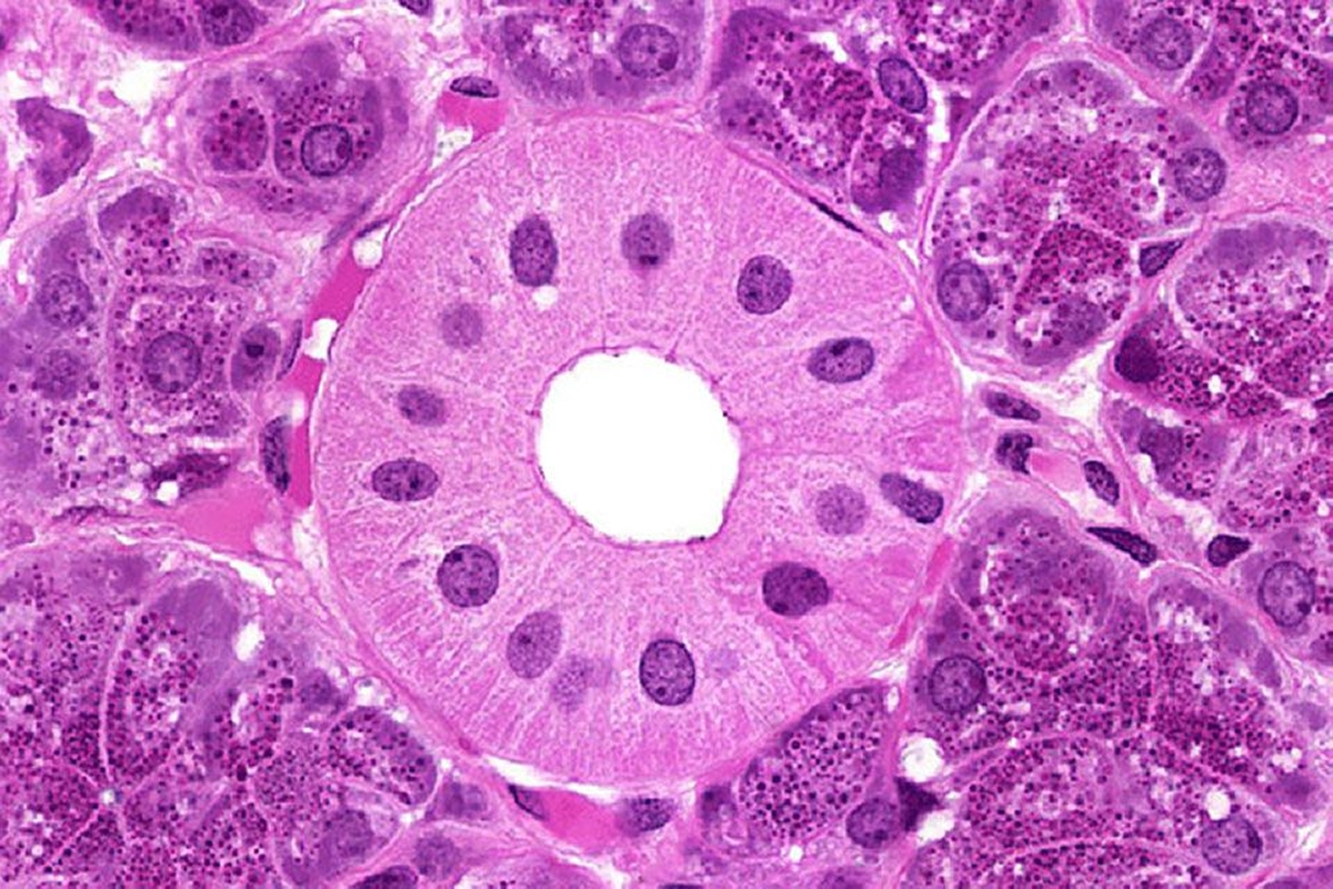
Histology Guide Virtual Microscopy Laboratory This gallery allows the browsing of the 307 microscope slides from the virtual slide box. cells and tissues. tissues are classified into four basic types: epithelium, connective tissue (includes cartilage, bone and blood), muscle, and nervous tissue. chapter 1. The atlas of human histology: a guide to microscopic structure of cells, tissues and organs by robert l. sorenson and t. clark brelje provides a print version of the core slides from this website. individual slides are presented as a series of images of increasing magnification to help convey a sense of scale and proportion. These purple (basophilic) structures are nuclei within the pink (eosinophilic) liver cell (hepatocyte). you can see the border of the nuclear membrane in these hepatocytes. additionally, you can see the cell membrane of the hepatocyte itself! zoom in through successive objectives until you reach 40x. zoom in on one nucleus. These lab worksheets are designed to guide you through a laboratory experience in histology. there are no labeled images here, as the goal is to get you to practice identifying structures on your own. i strongly suggest using a companion atlas or digital resource as you work through these exercises. i also suggest keeping a notebook (either.

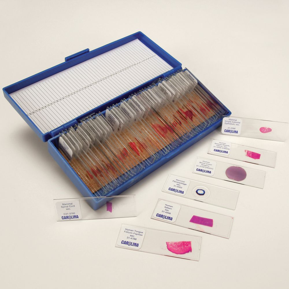
Basic Medical Histology Slide Set Carolina These purple (basophilic) structures are nuclei within the pink (eosinophilic) liver cell (hepatocyte). you can see the border of the nuclear membrane in these hepatocytes. additionally, you can see the cell membrane of the hepatocyte itself! zoom in through successive objectives until you reach 40x. zoom in on one nucleus. These lab worksheets are designed to guide you through a laboratory experience in histology. there are no labeled images here, as the goal is to get you to practice identifying structures on your own. i strongly suggest using a companion atlas or digital resource as you work through these exercises. i also suggest keeping a notebook (either. Slidebox. this website contains more than 250 virtual microscope slides for learning histology. cells and tissues. histology is organized into four basic types of tissue: (1) epithelium, (2) connective tissue (includes cartilage, bone and blood), (3) muscle, and (4) nervous tissue. Using this application makes you needless to the histology lab. besides, histological slides labeled and each labeled structure were described. also, each labeled structure is described by a simple schematic picture as well that makes learning much easier. you have to imagine the 3d structure of tissues by observing 2d sections of various.

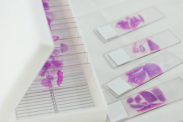
Best Histology Slides Stock Photos Pictures Royalty Free Images Istock Slidebox. this website contains more than 250 virtual microscope slides for learning histology. cells and tissues. histology is organized into four basic types of tissue: (1) epithelium, (2) connective tissue (includes cartilage, bone and blood), (3) muscle, and (4) nervous tissue. Using this application makes you needless to the histology lab. besides, histological slides labeled and each labeled structure were described. also, each labeled structure is described by a simple schematic picture as well that makes learning much easier. you have to imagine the 3d structure of tissues by observing 2d sections of various.

Comments are closed.