Full Form Of Opg In Dental X Ray Fullforms

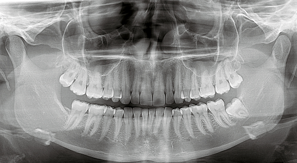
Full Form Of Opg In Dental X Ray Fullforms Orthopantomogram (opg) is a type of full mouth dental x ray scan that displays all the teeth of the upper and lower jaw. opg gives a panoramic or wide view of all the teeth and the surrounding bones of the face and jaw. it helps in diagnosing dental problems and assists in planning procedures such as implants and orthodontics. An orthopantomogram (opg) is an advanced type of full mouth dental x ray. an opg is a panoramic or wide view x ray, which displays all the teeth of the upper and lower jaw on a single film. it demonstrates the number, position and growth of all the teeth including those that have not yet surfaced or erupted. opg x ray produces a 2 dimensional.

What Is The Full Form Of Opg X Ray Leverage Edu During an opg the patient remains in a stationary position (seated or standing) while both the x ray source and film rotate in combination around the patient. the x ray source rotates from one side of the jaw, around the front of the patient, and then to the other side of the jaw. the film rotates opposite to the x ray source behind the patient. Opg (orthopantomogram) and lat ceph (lateral cephalometric radiograph) are special x rays of the lower face, teeth and jaws. an opg is a panoramic or wide view x ray of the lower face, which displays all the teeth of the upper and lower jaw on a single film. it demonstrates the number, position and growth of all the teeth including those that. In lateral cephalogram x ray, a radiographic image is a capture from the side view of the object. it gives similar details of all facial structures, bones and soft tissue as opg can generate but from a different view. it is commonly used by orthodontics to monitor the teeth alignment, the relation of teeth with the jaw, and open and crossbites. Pediatric dental emergency; tongue tie; sedation dentistry nitrous oxide sedation; laser dentistry; full mouth rehabilitation; invisalign provider; sleep airway dentistry; hypnodontics; digital dentistry; general dentistry. geriatric dentistry; whitening; orthodontic care; scaling & air polishing; tooth colored restorative fillings; root canal.

Opg X Ray Dental Scan Ltd In lateral cephalogram x ray, a radiographic image is a capture from the side view of the object. it gives similar details of all facial structures, bones and soft tissue as opg can generate but from a different view. it is commonly used by orthodontics to monitor the teeth alignment, the relation of teeth with the jaw, and open and crossbites. Pediatric dental emergency; tongue tie; sedation dentistry nitrous oxide sedation; laser dentistry; full mouth rehabilitation; invisalign provider; sleep airway dentistry; hypnodontics; digital dentistry; general dentistry. geriatric dentistry; whitening; orthodontic care; scaling & air polishing; tooth colored restorative fillings; root canal. An opg is an x ray of the lower face. like all x rays, it involves using short blasts of low level radiation to create images of inside the body – in this case, of the bones and teeth. the procedure for dental panoramic radiography consists of the patient resting their chin on a small shelf in front of the x ray machine and biting softly on a. Background. an orthopantomogram (opg) is a common radiograph used to identify the hard tissues of the oral cavity and surrounding skeletal structures. it is an extra oral radiograph that approximates the focal trough of the mandible. although resolution is not as detailed as intra oral radiographs for examination of the teeth, gross changes in.

Full Mouth X Ray Opg Shanti Dentals An opg is an x ray of the lower face. like all x rays, it involves using short blasts of low level radiation to create images of inside the body – in this case, of the bones and teeth. the procedure for dental panoramic radiography consists of the patient resting their chin on a small shelf in front of the x ray machine and biting softly on a. Background. an orthopantomogram (opg) is a common radiograph used to identify the hard tissues of the oral cavity and surrounding skeletal structures. it is an extra oral radiograph that approximates the focal trough of the mandible. although resolution is not as detailed as intra oral radiographs for examination of the teeth, gross changes in.

Comments are closed.