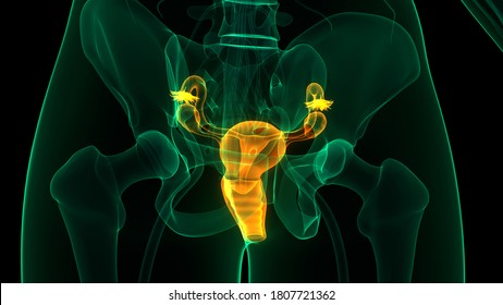
Female Reproductive System Labels Anatomy 3d Stock Illustration

Female Reproductive System Labels Anatomy 3d Stock Illustration In humans, the female reproductive system is immature at birth and develops to maturity at puberty to be able to produce gametes, and to carry a foetus to full term. the internal sex organs are the vagina, uterus, fallopian tubes, and ovaries. published 3 years ago. science & technology 3d models. reproduction. Anatomy explorer. the uterus and ovaries are the most vital organs of the female reproductive system. these organs work together to produce female sex hormones, produce and develop ova (egg cells), and support the development of a fetus during pregnancy. the uterus, also known as the womb, is a hollow, muscular, pear shaped organ found in the.

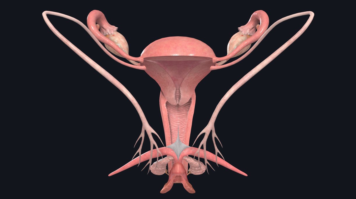
Female Reproductive System Labels Anatomy 3d Stock Illustration The internal genitalia include a three part system of ducts: the uterine tubes, the uterus, and the vagina. this system of ducts connects to the ovaries, the primary reproductive organs. the ovaries produce egg cells and release them for fertilization. fertilized eggs develop inside the uterus. 1. generating eggs: ovaries are the female gonads. The female reproductive system includes the ovaries, fallopian tubes, uterus, vagina, vulva, mammary glands and breasts. these organs are involved in the production and transportation of gametes and the production of sex hormones. the female reproductive system also facilitates the fertilization of ova by sperm and supports the development of. Learn about the anatomy and function of the vagina with innerbody's 3d illustrations. the vagina is an elastic, muscular tube connecting the cervix of the uterus to the vulva and exterior of the body. the vagina is located in the pelvic body cavity posterior to the urinary bladder and anterior to the rectum. measuring around 3 inches in length. Female anatomy includes the internal and external structures, including those responsible for hormones, reproduction, and sexual activity. the female reproductive system is essential for hormone regulation, sexual pleasure, pregnancy, breastfeeding, and more. the main parts of the female anatomy can be broken up into external and internal parts.

Female Reproductive System Labels Anatomy 3d Stock Illustration Learn about the anatomy and function of the vagina with innerbody's 3d illustrations. the vagina is an elastic, muscular tube connecting the cervix of the uterus to the vulva and exterior of the body. the vagina is located in the pelvic body cavity posterior to the urinary bladder and anterior to the rectum. measuring around 3 inches in length. Female anatomy includes the internal and external structures, including those responsible for hormones, reproduction, and sexual activity. the female reproductive system is essential for hormone regulation, sexual pleasure, pregnancy, breastfeeding, and more. the main parts of the female anatomy can be broken up into external and internal parts. 101 original medical illustrations have been created, presenting the anatomy of the female pelvis, including the pelvic bones (pelvimetry), the pelvic diaphragm, the urinary system (ureters, bladder and urethra), the female genital system (ovaries, uterine tubes, uterus, vagina, vulva and clitoris), the digestive system (rectum, anal canal and anus), as well as the pelvic blood supply, the. The major organs of the female reproductive system include: vagina: this muscular tube receives the penis during intercourse and through it a baby leaves the uterus during childbirth. uterus: this.

Female Reproductive System Anatomy 3d Stock Illustration 1 101 original medical illustrations have been created, presenting the anatomy of the female pelvis, including the pelvic bones (pelvimetry), the pelvic diaphragm, the urinary system (ureters, bladder and urethra), the female genital system (ovaries, uterine tubes, uterus, vagina, vulva and clitoris), the digestive system (rectum, anal canal and anus), as well as the pelvic blood supply, the. The major organs of the female reproductive system include: vagina: this muscular tube receives the penis during intercourse and through it a baby leaves the uterus during childbirth. uterus: this.

The Female Reproductive System Complete Anatomy

Comments are closed.