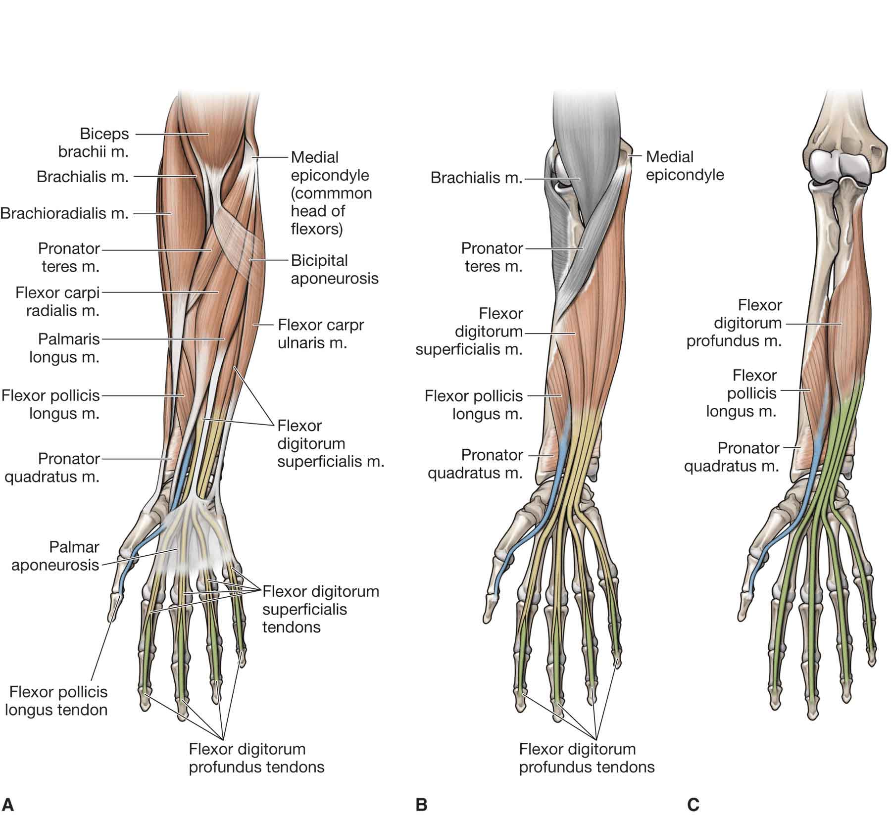
Diagram Of Forearm Muscles And Tendons

Forearm Diagram Muscles The elbow joint is a synovial joint that connects the arm and the forearm, providing 150 ْ of extension flexion movement. it consists of three joints; the humeroulnar joint, the humeroradial joint, and the proximal radioulnar joint, all within one articular capsule! the elbow joint is supported by three ligaments:. Lesson on the anatomy of the forearm: muscles and tendons.hey everyone! this is lesson 1 on the anatomy of the forearm. in this lesson, we look at the muscle.

Muscles Of The Anterior Forearm Superficial View Learn Muscles The inserting tendons of the 3 deepest muscles, the abductor pollicis longus, extensor pollicis brevis, and extensor pollicis longus, form the medial and lateral borders of the anatomical snuffbox — the triangular depression on the radial side of the dorsum, at the base of the thumb. these 3 muscles can together be referred to as the. Brachialis. this muscle lies underneath your biceps. it acts as a bridge between your humerus and ulna, one of the main bones of your forearm. it’s involved with the flexing of your forearm. The bicep tendon reflex tests spinal cord segment c6. coracobrachialis. the coracobrachialis muscle lies deep to the biceps brachii in the arm. attachments: originates from the coracoid process of the scapula. the muscle passes through the axilla, and attaches the medial side of the humeral shaft, at the level of the deltoid tubercle. The superficial muscles in the anterior compartment are the flexor carpi ulnaris, palmaris longus, flexor carpi radialis and pronator teres. they all originate from a common tendon, which arises from the medial epicondyle of the humerus. flexor carpi ulnaris. the flexor carpi ulnaris has two origins.

Forearm Anatomy Diagram The bicep tendon reflex tests spinal cord segment c6. coracobrachialis. the coracobrachialis muscle lies deep to the biceps brachii in the arm. attachments: originates from the coracoid process of the scapula. the muscle passes through the axilla, and attaches the medial side of the humeral shaft, at the level of the deltoid tubercle. The superficial muscles in the anterior compartment are the flexor carpi ulnaris, palmaris longus, flexor carpi radialis and pronator teres. they all originate from a common tendon, which arises from the medial epicondyle of the humerus. flexor carpi ulnaris. the flexor carpi ulnaris has two origins. Anatomy of the hand and wrist. your hands and wrists are a complicated network of bones, muscles, nerves, connective tissue and blood vessels. your hands and wrists help you interact with the world around you every day. talk to a healthcare provider if you have hand or wrist pain, especially if it’s getting worse over time. The muscle belly then crosses the entire upper arm and separates into two tendons. one tendons inserts onto the forearm bone, the radius, and the second spreads out to join the fascia along the upper part of the forearm. the tendons have 2 functions: to bend the elbow and to turn the palm of the hand towards the sky. triceps tendon.

Comments are closed.