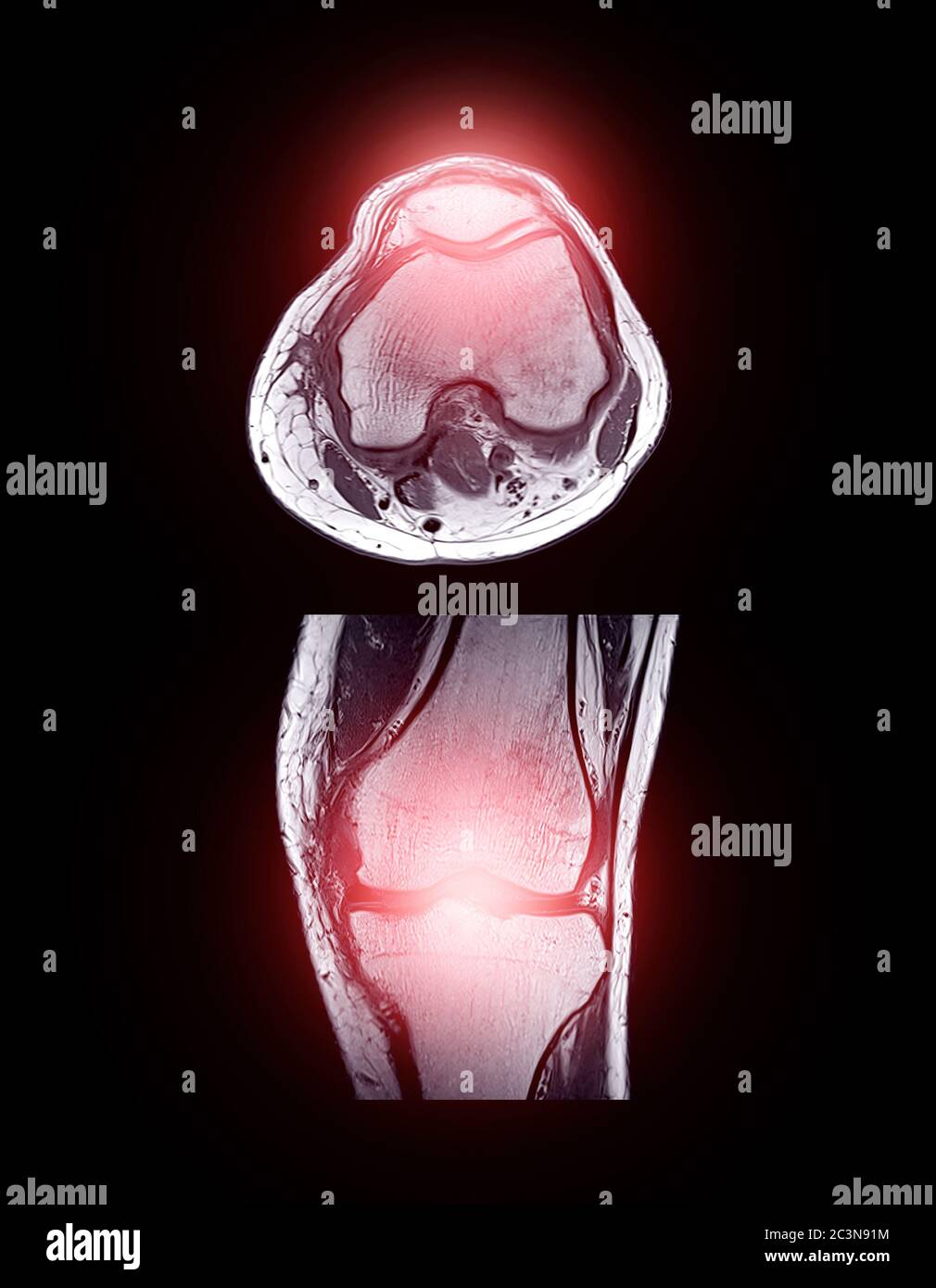
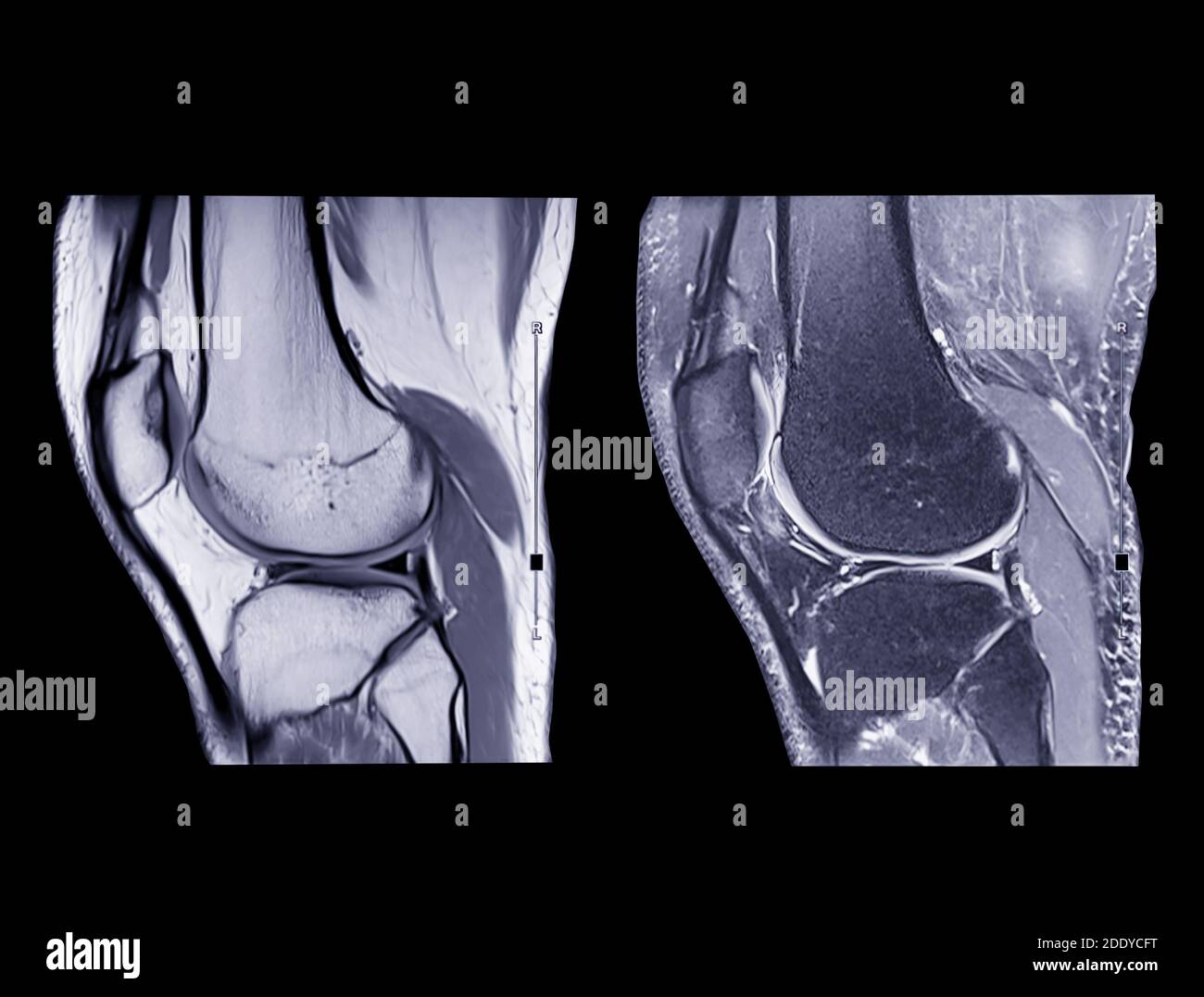
Compare Of Mri Knee Or Magnetic Resonance Imaging Of Knee Joi

Mri Knee Joint Or Magnetic Resonance Imaging Comparison Axial An The extent of knee damage can be evaluated clinically, but the development of magnetic resonance imaging (mri) allows for a more precise diagnosis of soft tissue and cartilage lesions in the knee. in the assessment of knee injuries, mri has proven to be a quick and non invasive imaging alternative to physical examination. Magnetic resonance imaging (mri) has been the gold standard for assessing knee cartilage thickness .however mri is expensive, not available to all patients at all times and not easily available for serial evaluation of cartilage status.

Magnetic Resonance Imaging Or Mri Knee Comparison Sagittal Pdw A This study supports the use of mri and clinical assessment in the diagnosis of chondral defects and internal knee derangement. clinical tests are reliable and have high sensitivity in diagnosing acl tears and chondral defects when compared to mri. not all lesions should routinely undergo mri for dia …. Accurate diagnosing of knee injuries is directly linked to taking the clinical history and making a careful physical examination. meniscal and ligament injuries of this joint can be evaluated by means of magnetic resonance imaging (mri) examinations, which provide images showing abnormalities of the morphology that are characterized. Abstract. knee pain is frequently seen in patients of all ages, with a wide range of possible aetiologies. magnetic resonance imaging (mri) of the knee is a common diagnostic examination performed for detecting and characterising acute and chronic internal derangement injuries of the knee and helps guide patient management. Magnetic resonance imaging (mri) of the knee joint has often been regarded as a noninvasive alternative to diagnostic arthroscopy. in day to day clinical practice, the mri scan is routinely used.

Magnetic Resonance Imaging Mri Knee Comparison Stock Photo 18601 Abstract. knee pain is frequently seen in patients of all ages, with a wide range of possible aetiologies. magnetic resonance imaging (mri) of the knee is a common diagnostic examination performed for detecting and characterising acute and chronic internal derangement injuries of the knee and helps guide patient management. Magnetic resonance imaging (mri) of the knee joint has often been regarded as a noninvasive alternative to diagnostic arthroscopy. in day to day clinical practice, the mri scan is routinely used. Signal to noise ratio. young adult. ultra high field mri at 7 t improved the overall diagnostic confidence in routine mri of the knee joint compared with that at 3 t. this is especially true for small joint structures and subtle lesions. higher spatial resolution was identified as the main reason for this improvement. To date, magnetic resonance imaging (mri) quantitative ultrasound assessment of medial meniscal extrusion defined by 2 mm threshold in patients with chronic knee pain in comparison to mri.

Comments are closed.