Anatomy Of The Ventricular Septal Defect In Congenita Vrogue Co

Congenital Heart Disease Septal Defects Ventricular S Vrogue Co Abstract. background: a ventricular septal defect (vsd) is an integral part of most congenital heart defects (chd). to determine the prevalence of vsd in various types of chd and the distribution of their anatomic types. methods: we reviewed 1178 heart specimens with chd from the anatomic collection of the french reference centre for complex. Abnormal completion of this structure during embryonic and fetal life results in a ventricular septal defect (vsd). isolated vsds are the most frequent congenital heart defects and are also an integral part of more complex cardiac anomalies [1–3]. it is thus the most common phenotype in all congenital heart defects.

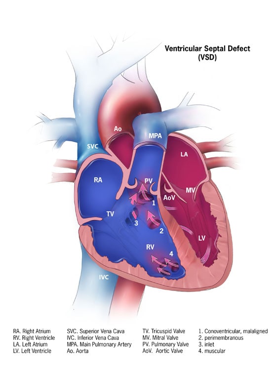
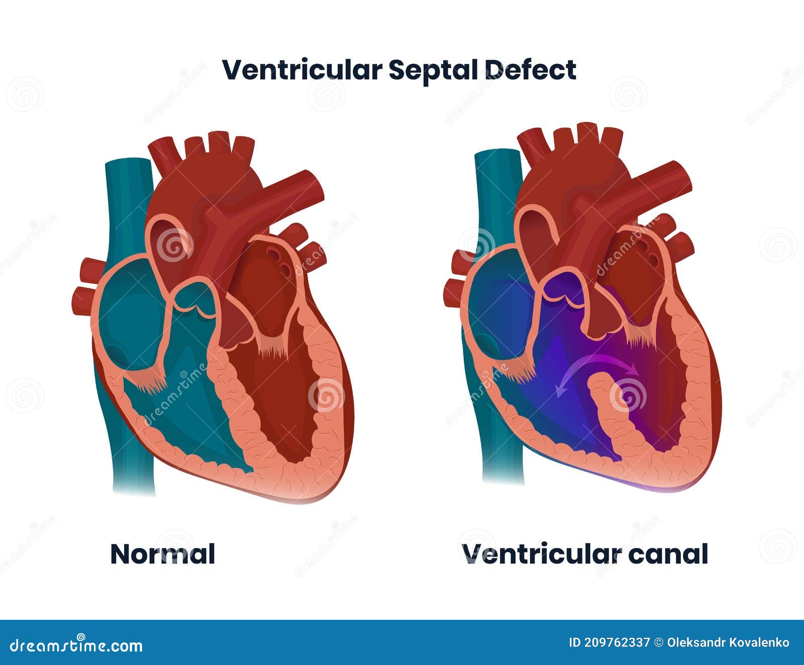
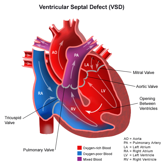
Anatomy Of The Ventricular Septal Defect In Congenita Vrogue Co Ventricular septal defect (vsd) is the most prevalent congenital heart disease (chd) and is easily misdiagnosed or missed [1][2] [3] [4]. it can exist alone (hereafter referred to as an isolated. The anatomic distribution of vsd is similar in isolated vsd, coa and tga, while the v sd is predominantly outlet in outflow tract defects except tga which reinforces the allegedly different mechanisms in tga and cardiac neural crest defects. backgrounda ventricular septal defect (vsd) is an integral part of most congenital heart defects (chd). to determine the prevalence of vsd in various. Ventricular septal defect (see figure ventricular septal defect) is the 2nd most common congenital heart anomaly after bicuspid aortic valve, accounting for 20% of all defects. it can occur alone or with other congenital anomalies (eg, tetralogy of fallot , complete atrioventricular septal defects , transposition of the great arteries ). Ventricular septal defects (vsds) are the most common congenital cardiac anomaly in children and are the second most common congenital abnormality in adults, surpassed only by a bicuspid aortic valve. the primary pathophysiology involves an abnormal communication between the right and left ventricles, leading to shunt formation and subsequent hemodynamic compromise in vsd. while spontaneous.

Ventricular Septal Defect Congenital Heart Disease Co Vrogue Ventricular septal defect (see figure ventricular septal defect) is the 2nd most common congenital heart anomaly after bicuspid aortic valve, accounting for 20% of all defects. it can occur alone or with other congenital anomalies (eg, tetralogy of fallot , complete atrioventricular septal defects , transposition of the great arteries ). Ventricular septal defects (vsds) are the most common congenital cardiac anomaly in children and are the second most common congenital abnormality in adults, surpassed only by a bicuspid aortic valve. the primary pathophysiology involves an abnormal communication between the right and left ventricles, leading to shunt formation and subsequent hemodynamic compromise in vsd. while spontaneous. Abstract. congenital ventricular septal defects (vsd) are the second most common congenital heart defect after bicuspid aortic valves. congenital vsds vary greatly in location, clinical presentation, and associated lesions. a full understanding of these defects requires a certain familiarity with the septation of the ventricles and normal post. Van praagh r, geva t, kreutzer j. ventricular septal defects: how shall we describe, name and classify them? j am coll cardiol 1989; 14:1298. rudolph am. ventricular septal defect. in: congenital diseases of the heart: clinical physiological considerations, rudolph am (ed), futura publishing company, new york 2001. p.197. moe dg, guntheroth wg.

Anatomy Of The Ventricular Septal Defect In Congenita Vrogue Co Abstract. congenital ventricular septal defects (vsd) are the second most common congenital heart defect after bicuspid aortic valves. congenital vsds vary greatly in location, clinical presentation, and associated lesions. a full understanding of these defects requires a certain familiarity with the septation of the ventricles and normal post. Van praagh r, geva t, kreutzer j. ventricular septal defects: how shall we describe, name and classify them? j am coll cardiol 1989; 14:1298. rudolph am. ventricular septal defect. in: congenital diseases of the heart: clinical physiological considerations, rudolph am (ed), futura publishing company, new york 2001. p.197. moe dg, guntheroth wg.

Ventricular Septal Defect Vsd Classification Pathophy Vrogue Co

Comments are closed.