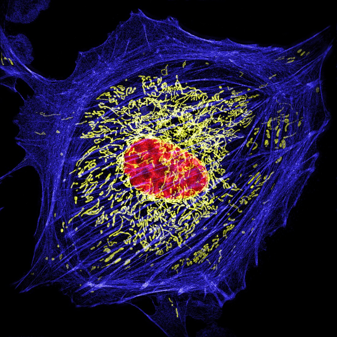
An Introduction To Super Resolution Microscopy Of Living Cells

New Fluorescent Probes To Image Live Cells With Super Resolution Super resolution microscopy is a rapidly developing field that allows biologists to 'see' objects in living cells much smaller than previously thought possib. The introduction of a. y. fluorescent probes for super resolution imaging in living cells. nat. rev. g. et al. a near infrared fluorophore for live cell super resolution microscopy of.

Optics Photonics News Super Resolution Microscope Expands View Of Fluorescence microscopy allows biologists to selectively label and observe cellular components with high sensitivity in fixed and living samples (combs and shroff, 2017; kremers et al., 2010; masters, 2010), and is one of the leading technologies used to drive discoveries in life sciences. Despite this, the application of super resolution microscopy to dynamic, living samples has thus far been limited and often requires specialised, complex hardware. here we demonstrate how a novel analytical approach, super resolution radial fluctuations (srrf), is able to make live cell super resolution microscopy accessible to a wider range of. In order to penetrate further into living cells, two photon microscopy can be combined with sted (moneron and hell, 2009), allowing super resolution live cell data to be taken up to 30 μm into tissue (takasaki et al., 2013). however, when attempting to achieve super resolution at these depths, the problem of aberration has to be considered. The super resolution techniques in biological research are also introduced. key words: super resolution microscopy, sted, palm, storm, sim, sax introduction optical microscopy has played a key role in biological and medical fields since optical microscopy allows us to image and investigate microorganisms, cells, tissues and organs in living.

Sim Hms Super Resolution Microscopy In The Department Of Cell In order to penetrate further into living cells, two photon microscopy can be combined with sted (moneron and hell, 2009), allowing super resolution live cell data to be taken up to 30 μm into tissue (takasaki et al., 2013). however, when attempting to achieve super resolution at these depths, the problem of aberration has to be considered. The super resolution techniques in biological research are also introduced. key words: super resolution microscopy, sted, palm, storm, sim, sax introduction optical microscopy has played a key role in biological and medical fields since optical microscopy allows us to image and investigate microorganisms, cells, tissues and organs in living. Super resolution fluorescence microscopy has transformed understanding of the structure and function of many biological systems. however, challenges are still present, and to maximize the impact of super resolution microscopy, further technological advancements are still needed. fig. 4. Summary. since its initial demonstration in 2000, far field super resolution light microscopy has undergone tremendous technological developments. in parallel, these developments have opened a new window into visualizing the inner life of cells at unprecedented levels of detail. here, we review the technical details behind the most common.

Comments are closed.